
Product.
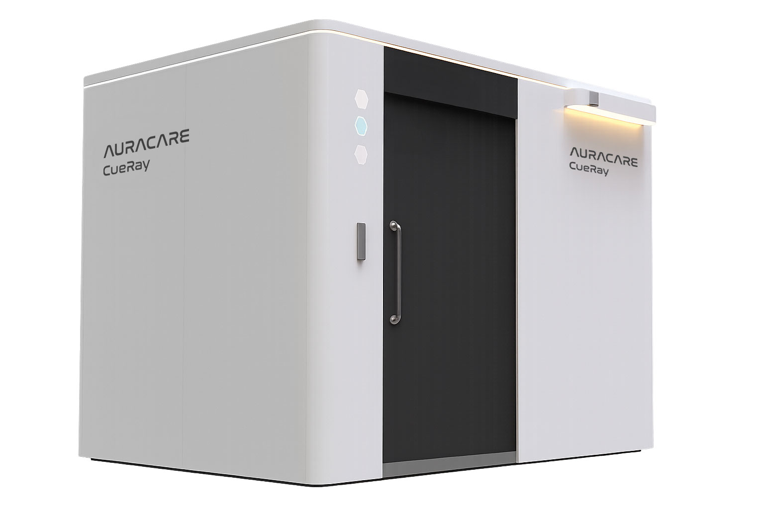
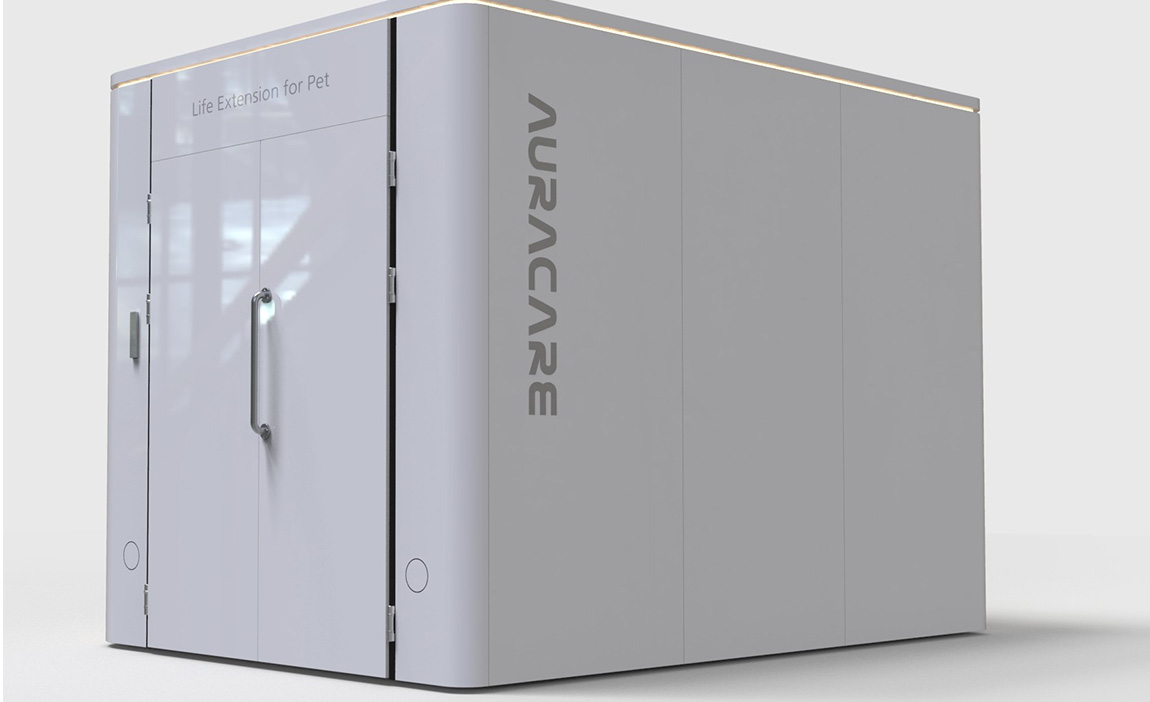
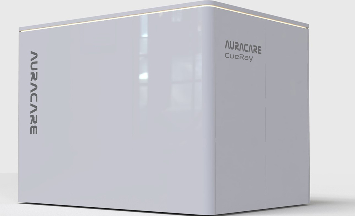
CueRay
Feature
-
1World’s first radiation cancer treatment device for companion animals
-
2Radiation treatment with image-guided alignment using a single radiation source.
-
3Minimization/optimization of device through optimal design for pets.
-
4Radiation shielding solutions that require no changes to existing building structures.
-
5Strong partnerships across universities, research institutions, hospitals, and industry to advance veterinary radiation therapy.
-
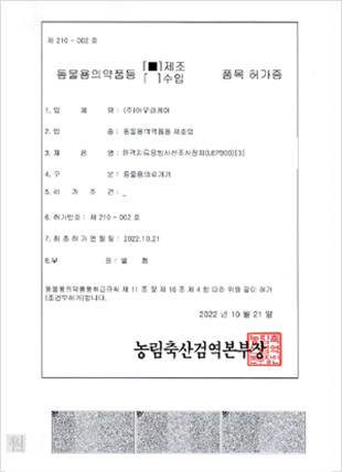
6Approved by the Animal and Plant Quarantine Agency (APQA)
Licensed as a veterinary medical device (License No. 210–002).
Specification
| Items | CueRay3 | CueRay7 | ||
|---|---|---|---|---|
| Electrical | Input Voltage | 100~220Vac, Single Phase , 50/60hz, 8kW | 100~220 Vac, Single Phase , 50/60hz, 8kW | |
| Mechanical | Dimension(W*L*H) | 240cm * 323cm * 240cm | 240cm * 323cm * 240cm | |
| (with Self-shielding Cabinet) | (with Self-shielding Cabinet) | |||
| Total Weight | about 8,000kg | about 8,000kg | ||
| (with Self-shielding Cabinet) | (with Self-shielding Cabinet) | |||
| Gantry Rotation Range | ± 180° | ± 180° | ||
| Collimator Rotation Range | ± 90° | ± 90° | ||
| Couch Laterial Range(X) | ± 9cm | ± 9cm | ||
| Couch Longitudinal Range(Y) | 0 to 70cm | 0 to 70cm | ||
| Couch Vertical Range(Z) | 0 to -30cm (from ISO Center) | 0 to -30cm (from ISO Center) | ||
| X-ray | X-ray Tube(2 Modes) | Treatment Mode : Max 14mA at 300kV | Treatment Mode : Max 14mA at 300kV | |
| Image Mode : Max 25mA at 100kV | Image Mode : Max 25mA at 100kV | |||
| SSD | 60cm | 60cm | ||
| Max. Field Size | Imaging Mode | 15cm * 15cm | 15cm * 15cm | |
| Treatment Mode | 15cm * 15cm | 15cm * 15cm | ||
| (Divided by 3 segments of 5cm*15cm) | ||||
| MLC | 26 pairs manual leaf | 14 pairs automatic leaf | ||
| - 20 * 2 leaves, 0.5cm | - 10 * 2 leaves, 0.5cm | |||
| - 6 * 2 leaves, 1cm | - 2 *2 leaves, 2.5cm | |||
| - 2 *2 leaves, 5cm | ||||
| Output | 1.07Gy/min at SSD 60cm | 1.07Gy/min at SSD 60cm | ||
| Patient Capacity | width ≤30cm, Weight ≤40kg | width ≤30cm, Weight ≤40kg | ||
Hardware
-
 Self-Shielding Cabinet
 -
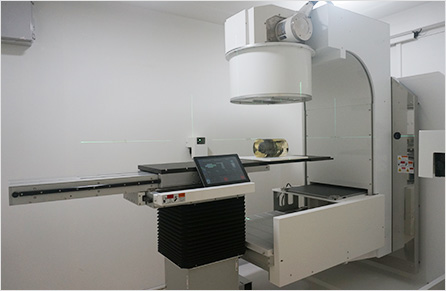 CueRay Main
 -
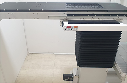 Couch
Couch -
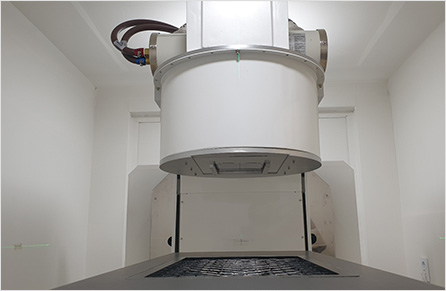 Head unit
 -
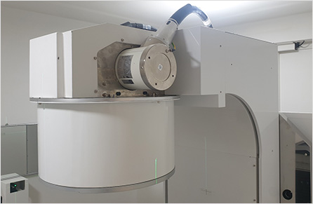 X-ray tube
 -
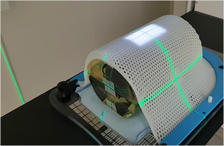 Phantom Positioning
Software
AuraConsole
CueRay SW
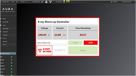
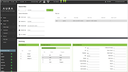
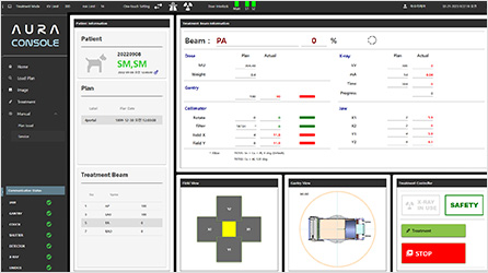
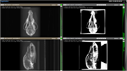
X-ray Warm Up UI
AuraRemote
CueRay remote control SW with Tablet PC
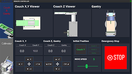
AuraRemote
AuraVet
Patient & treatment information management SW

AuraRemote
Performance Test
Tests for mechanical properties
| Item | Range of Operation | Specified Tolerance | Measured Error |
|---|---|---|---|
| Gantry Rotation | 360 ° | ± 2° | -0.5° |
| Collimator Rotation | 180° | ± 2° | 0.1° |
| Target-to-Isocenter Distance (TID) | 60 mm | ± 2 mm | 0.3 mm |
| Couch Vertical Range (Z axis) |
300 mm | ± 2 mm | 1.0 mm |
| Couch Vertical Range (X axis) |
±90 mm | ± 2 mm | 0.1 mm |
| Couch Vertical Range (Y axis) |
700 mm | ± 2 mm | 0.1 mm |
| Radiation Field Size | 15 x 15 cm | ± 2 mm | 0.6 mm |
Starshot Test
-
 Collimator Starshot Test
 -
Gantry Starshot Test
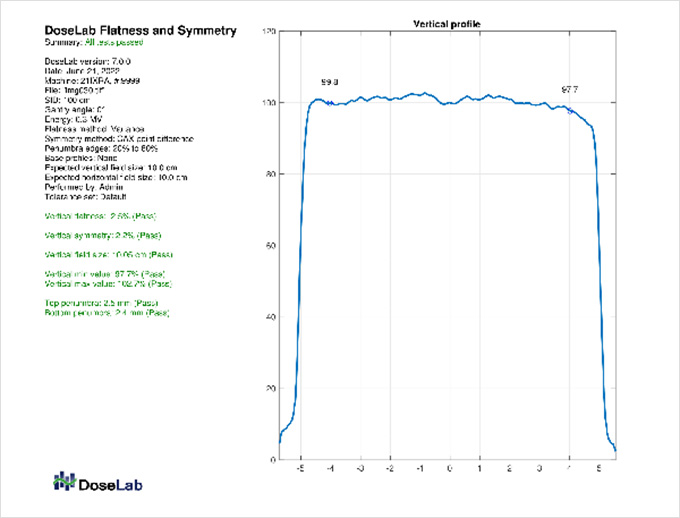
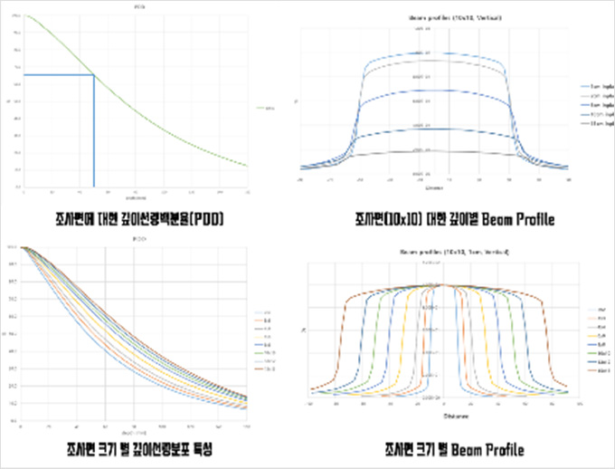
Beam Parameters
Certifications
-
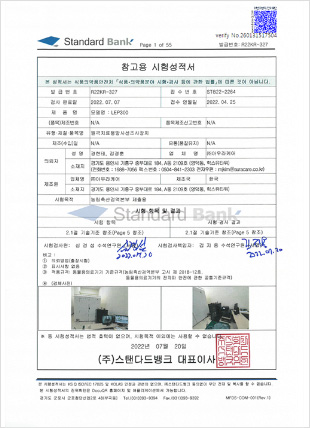 Electrical and Mechanical Safety &
Manufacturer’s Performance Test Report -
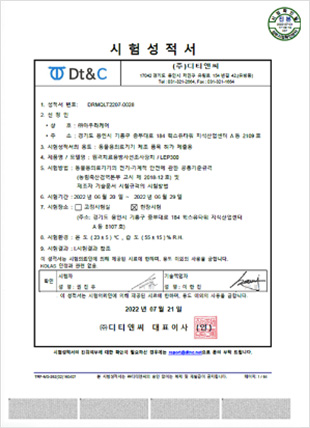 Electrical and Mechanical Safety &
Manufacturer’s Performance Test Report -
 Collimator Starshot Test
Case Study
-
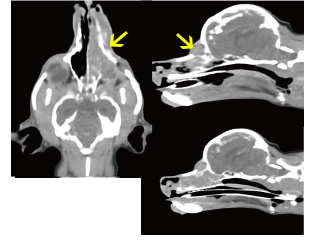 1Simulation CTBaseline tumor dimensions prior
to radiation therapy initiation -
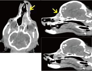 23 Weeks Post-RTInitial volume reduction of 19%
observed. -
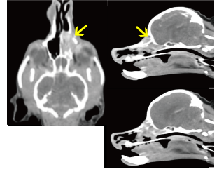 37 Weeks Post-RTFurther reduction to 49% of
original volume.
▪ Sequential CT imaging demonstrates progressive tumor response to radiation therapy over time.
-
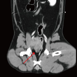 1Pre-Treatment CTLarge 4cm mass in right retroperitoneal space
with proximity to critical vascular structures -
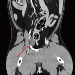 27 Weeks Post-RTDramatic reduction in tumor size creating
potential surgical window
Protocol: Total 32Gy, 8Gy × 4Fr
This case demonstrates the value of radiation therapy for cytoreduction of inoperable or difficult-to-access tumors.
-
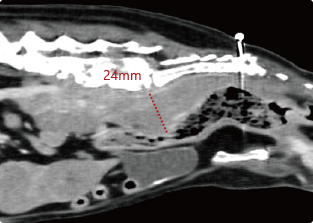
Simulation CTD+57
-
2weeks after 2nd RTD+93

CT imaging two weeks after RT completion showed reduction in sublumbar lymph node size and decreased compression of descending colon. This demonstrates palliative benefit of radiation therapy even in cases with distant metastasis.
-
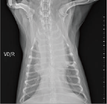 Initial Presentation (July 12, 2024)Prominent cranial mediastinal mass with associated pleural effusion. Note the increased opacity in the cranial thorax.
 -
7 Days After Single RT SessionMarked reduction in mediastinal width and density. Improved thoracic radiographic appearance with reduction in pleural fluid.
Thymomas in veterinary patients often show excellent radiosensitivity, with significant improvement possible after minimal radiation exposure. This response pattern is similar to that observed in human patients.

